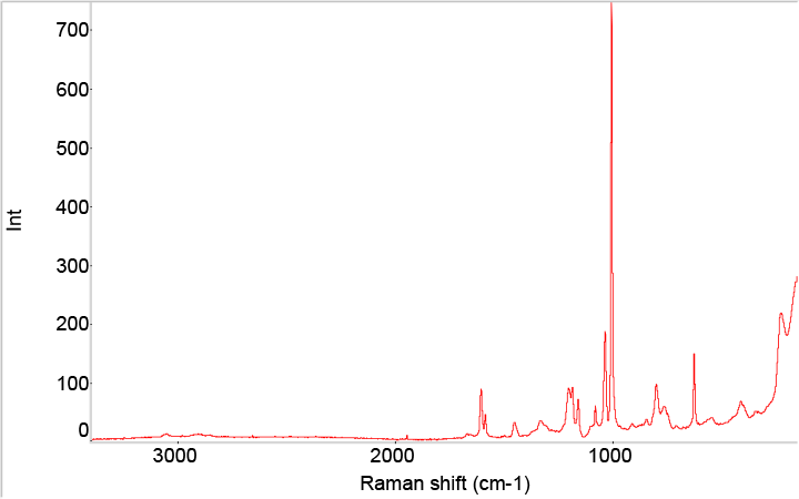
Confocal Raman microspectroscopy unites the spectral information from Raman spectroscopy with the spatial resolution of a confocal light microscope to provide high-resolution chemical imaging. The confocal optics of the light microscope enable the analysis volume within the sample to be spatially discrete with high resolution in both the lateral (XY) and depth or axial (Z) axes. The synergism between the spectral and spatial information enables the chemical analysis of individual particles, discrete sample features, or thin layers/films down to less than 1 micrometer in size and in some cases down to 100 nanometers. When contaminants or unknown particles are found in a sample, the high spatial resolution oftentimes allows for in-situ analysis without further isolation, as with commonly encountered particles embedded in the coating of pharmaceutical tablets.
Improved Pharma has expertise in both spectral interpretation using vibrational fundamental modes and comparison against our Raman spectral libraries. Our in-house spectral libraries contain several thousand compounds, which we use to identify unknown particles or characterize the properties of materials. Our confocal Raman microscope is equipped with two lasers (785 nm and 532 nm), a fully automated stage for mapping, and a selection of microscope objectives for specialized analyses.
Improved Pharma’s instrument is a HORIBA XploRA™ PLUS Confocal Raman Microscope. A total of four grating options are available.
To learn more about how Raman microspectroscopy can be used to solve pharmaceutical research challenges, please refer to these recent blog posts:

